Researchers at UC San Diego have developed a wearable, self-operating ultrasound cardiac imaging device that can provide information about the heart continuously for at least 24 hours, even during exercise. Traditional hand-held ultrasound devices used to take images of the heart, like during echocardiograms, are commonly used in hospitals by clinicians to assess a patient’s cardiovascular system while they lie down. These researchers hope this device will replace this traditional imager.
“Cardiac ultrasound imaging is one of the primary tools physicians have for easily and noninvasively evaluating the status of a patient’s heart,” nanoengineering doctoral student Ray Wu, who participated in designing the device layout, wrote in an email to The UCSD Guardian. “Cardiac imaging can reveal abnormalities in the heart’s structure or motion that would indicate disease.”
The ultrasound imager they have developed greatly differs from those commonly used in hospitals. Wu explained that traditional cardiac ultrasound imagers are bulky, static, and require clinicians to steadily, carefully operate a probe and interpret its images. Theirs, however, is small, wearable, and can interpret images on its own.
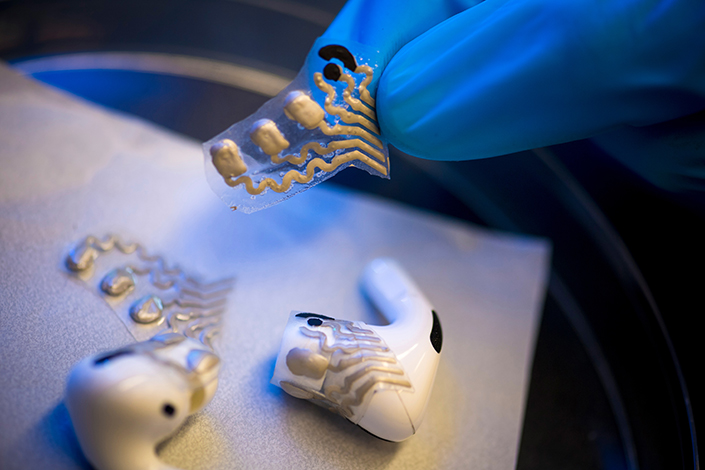
“This device takes the capabilities of a traditional ultrasound imager and packages it into a thin and small wearable device the size of a postage stamp [which] can adhere to the skin on its own and interpret the images automatically using a deep learning algorithm, removing much of the need for a trained operator,” Wu said.
These trained operators are not always a resource available in every hospital, making this gadget particularly valuable in underfunded areas, remote regions, and Global South countries, Principal Investigator Dr. Sheng Xu explained.
The device has additional valuable capabilities; it can also provide continuous imaging for more than a day. Nanoengineering doctoral student Hao Huang, who participated in fabricating the device and developing its image processing ability, added that it may be possible to wear the device for longer.
“In principle, wearing the device for over 24 hours is generally achievable. The maximum wearing duration is not determined now, but we expect that it can be worn for more than one week,” Huang said.
For this reason, the device delivers more information about cardiac activity than traditional imagers, helping physicians to diagnose more accurately, according to nanoengineering postdoctoral researcher Dr. Hongjie Hu. A device can only provide limited data if a patient is required to stay still, and might mean that clinicians miss possible problems with the cardiovascular system in that particular moment. Since the device can be used while in motion, Hu said, it provides a more diverse data set.
“Potential applications include continuously monitoring the heart in daily life, during exercise, during surgery, and much more,” Wu explained. “This will open up the possibility to detect previously undetectable symptoms of disease, identify symptoms in their very early stages, and greatly improve patient outcomes.”
Their device is especially pertinent because of the sheer number of people it may assist. According to the American Heart Association, 121.5 million adults in the United States, almost 50%, have cardiovascular disease. And the Centers for Disease Control and Prevention reports that heart disease is the leading cause of death in the U.S.
“After learning in a casual chat that one of our colleague’s relatives had died of a sudden heart attack, we realized our technology could make real impacts on people’s lives by continuously monitoring cardiac functions to reduce the mortality rate,” Huang said.
He explained the science behind the device; it works by transmitting and receiving ultrasound waves through a particular material chosen as a transducer, which converts energy from one form to another. The material was chosen for its ability to produce stronger signals than others.
“Piezoelectric [material] can turn mechanical energy into electrical energy,” Hu explained. “We gave the material [an electrical] pulse … After getting the voltage, the material vibrates to generate an ultrasound wave, which goes inside the human body. When the ultrasound meets the different tissues, it bounces back because the tissues have different acoustic impedance.”
Still, Hu admitted that while the device is innovative and can change the scope of cardiac imaging as it stands today, he hopes to continue to develop the technology even further.
“Our device is still tethered by flexible cables; it’s not wireless …. We still need to improve … the size of the backhand system, to integrate the device. [Then,] you could wear it, for example, while swimming.”
Nevertheless, he does not deny how much it can help clinicians and patients, and he hopes it will have a positive impact on the field of medicine as a whole.
“We specifically targeted the heart, because some cardiac diseases happen suddenly,” Hu said. “There’s no prediction you can make to know [when it] will happen … by traditional ultrasound. But using our device, if you wear it for a long time, it can capture the onset of this disease. I think that can greatly influence the medical field.”
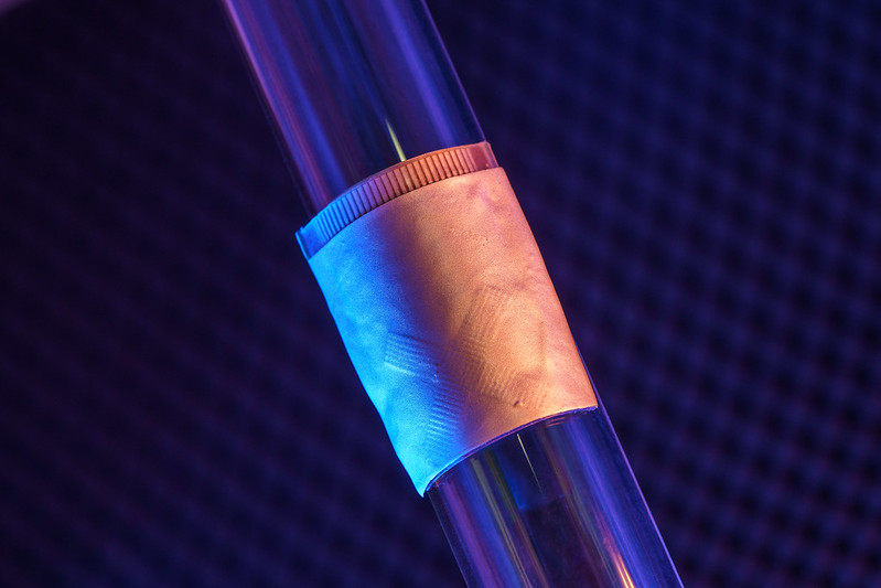
Photo courtesy of David Baillot for UC San Diego Jacobs School of Engineering